Published by: Dr. Rosa Ma. Montezuma : January 22, 2026.
Updated by: Dr. Miguel Ángel S.: June 11, 2026.
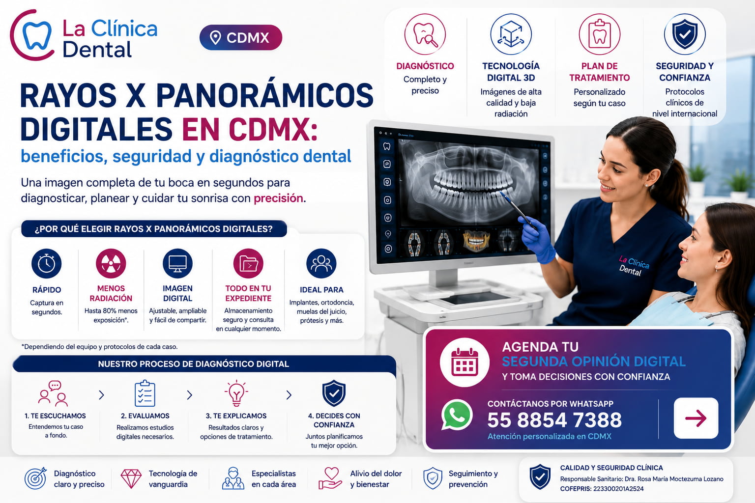
Digital panoramic X-rays are 2D dental X-rays that show teeth, jaws, joints, and bone structures in a single image. At La Clínica Dental CDMX, they are used as part of the 360 digital dental diagnosis to evaluate orthodontics, wisdom teeth, implants, prosthetics, and oral rehabilitation, and to determine when it is necessary to supplement the diagnosis with advanced dental technology such as 3D CBCT or an intraoral scanner.
The main advantage over conventional systems is that the image is obtained quickly, digitally, and is easy to analyze on screen. The decision to take an X-ray should be made on a case-by-case basis: an X-ray is not taken “routinely,” but only when it provides useful information for diagnosing, planning, or monitoring dental treatment.
Are you looking for a dental evaluation with digital X-rays in Mexico City?
At La Clínica Dental, we can combine digital panoramic X-rays with clinical evaluation, specialist consultations, and additional tests as needed for your specific case. This allows us to develop a clear treatment plan for orthodontics, implants, wisdom teeth, dentures, or comprehensive dental restoration.

What are digital panoramic X-rays?
How do they work?
Instead of film, a digital sensor or a special plate is used to capture the image.
The information is sent to a computer and processed with specific software.
The radiograph is viewed directly on a screen a few seconds after it is taken.
Main benefits
Lower radiation exposure (in many systems)
Many modern digital devices are designed to use lower radiation doses than analog systems, while maintaining adequate diagnostic quality.
Immediate image
No need to develop the film: the image appears on the computer almost instantly.
This speeds up the consultation and allows decisions to be made at the same time.
Image adjustments
You can modify brightness, contrast, zoom in, and highlight areas of interest.
This reduces the need to repeat the shot due to exposure problems.
Storage and backup
Radiographs can be stored in the patient’s digital record.
It is easier to organize, search, and compare images over time.
Backups can be made so you don’t lose the information.
Ease of sharing
They can be sent electronically to other specialists or to the patient.
Ideal for second opinions, interconsultations, or referrals.
Lower environmental impact
No developing chemicals or disposable films are used.
Reduces the consumption of paper and plastic associated with plates and envelopes.
Where can I get digital panoramic X-rays in Mexico City?
Ideally, a digital panoramic X-ray should be taken at a dental clinic that can interpret the results as part of a comprehensive treatment plan, rather than simply providing an isolated image. At La Clínica Dental CDMX, a digital panoramic X-ray can be part of a comprehensive evaluation to detect impacted wisdom teeth, bone loss, infections, root abnormalities, bite problems, or the need for treatment by specialists.
When the case requires greater precision, the panoramic X-ray can be supplemented with 3D CBCT dental tomography, an intraoral scanner, and digital design. This workflow is especially useful before beginning dental implants, orthodontic treatment, maxillofacial surgery, or comprehensive oral rehabilitation.
Conventional (analog) panoramic X-rays
How do they work?
A radiographic film is used (similar to old cinema or photography).
After exposure to X-rays, the film is developed in a dark room with chemicals.
The image is displayed on a physical plate that is placed in front of a negatoscope (light box).
Advantages
Technology proven for decades.
Initially more economical equipment (historically).
Disadvantages
Use of chemicals (developer and fixer), which require proper handling and disposal.
Longer time between shooting and viewing (the film has to be developed).
Difficulty adjusting brightness or contrast: if the shot is too light or dark, it must be repeated.
Physical preservation of the plates (they deteriorate, take up space, can be lost).
Less ease in sharing the image (the plate must be scanned or transported).
Digital vs. Conventional Panoramic X-rays: Key Differences
| Appearance | Digital panoramic | Conventional panoramic |
|---|---|---|
| Image Capture | Uses a sensor or digital plate to generate the image on the screen. | Uses radiographic film that requires chemical development. |
| Display Time | The image appears almost immediately and can be reviewed during the appointment. | You must wait for the physical development before analyzing the film. |
| Image Adjustment | Allows you to adjust brightness, contrast, and zoom to better explain the case. | Physical images offer less flexibility for diagnostic adjustments. |
| Radiation | In many digital systems, radiation exposure may be lower, depending on the equipment, protocol, and clinical indication. | It may require a higher dose or a repeat exposure if the image is poorly exposed. |
| Digital Medical Record | It is integrated into the patient’s medical record and can be shared for consultations or a second opinion. | The physical film may deteriorate, be lost, or require scanning. |
| Environmental Impact | It requires no developing chemicals or physical film. | It uses developer, fixer, and physical materials that require proper handling. |
When is a panoramic X-ray sufficient, and when is a 3D CBCT scan needed?
A digital panoramic X-ray provides an excellent initial overview, but it does not replace all dental imaging studies. In complex cases, the dentist may request additional images to confirm details that the 2D panoramic X-ray does not show in sufficient detail.
| Study | What Shows It Best | When it is indicated |
|---|---|---|
| Digital Panoramic X-ray | Overview of teeth, jaws, joints, and bone structures. | Initial evaluation, orthodontics, wisdom teeth, prosthetics, comprehensive checkup. |
| Periapical X-ray | Close-up of a tooth, root, and the area around the apex. | Localized pain, deep cavity, root canal, follow-up on a specific tooth. |
| 3D CBCT Scan | Bone volume, thickness, height, and relationship to nerves and maxillary sinuses. | Implants, guided surgery, bone loss, complex cases, or surgical planning. |
| Intraoral Scanner | Digital impression of teeth and bite without traditional molds. | Aligners, crowns, veneers, smile design, mouthguards, and cosmetic restoration. |
If your goal is to get implants, have a crown checked, or confirm whether a previous treatment plan is correct, request a digital dental second opinion in Mexico City. If you already know you need extensive treatment, find out how to organize a phased dental treatment plan.
How much radiation is involved in a digital panoramic X-ray?
The radiation dose from a panoramic dental X-ray is usually low, but it can vary depending on the equipment, the settings, the patient’s age, and the clinical purpose of the exam. Therefore, the proper approach is not to take X-rays routinely, but to order them when they help answer a diagnostic question or plan treatment.
At La Clínica Dental, an X-ray is ordered when it provides useful information for your case: for example, to evaluate wisdom teeth, analyze bone loss, plan orthodontic treatment, check on implants, or design a dental restoration. If you have a confirmed pregnancy or suspect you might be pregnant, have relevant medical history, or have had recent tests, please let us know before the procedure so the clinical team can determine the appropriate protocol.
Panoramic vs. Periapical: The Complete Map vs. the Magnifying Glass
If the panoramic radiograph is the complete map of your mouth, the smaller radiographs, called periapical or bitewing, are the magnifying glass. While the panoramic offers a dental image of the entire mouth to assess the general structure, the periapical focuses on a tiny area with extraordinary sharpness.
Surely you remember them: they are those small plates that you hold with your teeth inside your mouth for a few seconds. Its superpower is detail. They are irreplaceable for an accurate dental diagnosis of problems such as a cavity between two teeth or to examine the tip of the root of a tooth that causes pain. The panoramic tells you where to look, and the periapical shows you exactly what is happening there.
Therefore, it is not a competition of panoramic vs. periapical radiograph, but a teamwork. Your dentist uses the map to orient themselves and the magnifying glass to investigate. But when the problem is more complex and you need to see the depth and volume, the technology offers a three-dimensional view.
When a 3D View is Needed: A Brief Look at CBCT Tomography
Sometimes, a flat map is not enough. To place a dental implant, the dentist needs to know how thick and dense the available bone is. A 2D image is like a photo of a wall: it shows the height and width, but not its thickness. For that, a third dimension is needed.
For these cases, there is cone beam computed tomography (CBCT). Although the name sounds imposing, its function is simple: it is a 3D scanner for the mouth. The machine rotates around you capturing hundreds of images that a computer combines to create an exact three-dimensional model of your teeth and jaw. It is the quintessential tool for dental implant preparation.
With this 3D model, the specialist can virtually navigate through your anatomy, measure spaces with millimeter precision, dodge nerves, and ensure that the implant will have perfect support. It is also essential for an evaluation of the temporomandibular joint in complex cases. It is not a routine study, but when maximum precision is necessary, this technology guarantees the safety and success of the treatment.
What You Should Know About the Price of a Dental Panoramic Radiograph?
When talking about medical tests, it is logical to think about the cost. The price of a dental panoramic radiograph may vary, but it is crucial to understand it not as an expense, but as an investment in dental health. This image is the basis on which your dentist will build an accurate diagnosis. Without it, it is like asking an architect to design a house without knowing the land.
The true value of the panoramic is its ability to detect hidden problems before they worsen. Identifying a poorly positioned wisdom tooth in time can avoid emergency surgery, and seeing the arrangement of all the roots is essential for a successful orthodontic treatment. Therefore, the cost of orthopantomography saves you money and complications in the long term.
In addition, in many cases this study is already part of a comprehensive plan for orthodontics, implants, or wisdom teeth extraction. Often, the price is included in the total treatment budget, not as an additional surprise.
What should a patient know before having a panoramic X-ray?
Digital panoramic X-rays are quick, non-invasive, and painless. Their use should be based on a specific clinical need: evaluating wisdom teeth, assessing bone loss, planning orthodontic treatment, evaluating implants, or studying a case of oral rehabilitation.
Exposure is usually low, but it can vary depending on the equipment, settings, and protocol used. Therefore, the proper approach is to perform the test only when it provides useful information for diagnosing, planning, or monitoring treatment.
In which treatments are digital panoramics especially useful?
Digital panoramic X-rays are very useful in:
Orthodontics: evaluation of roots, tooth eruption, absence or excess of teeth, wisdom teeth.
Dental implants (as a first approximation): to see spaces, approximate bone height, missing teeth.
Oral surgery: wisdom teeth, cysts, lesions.
Pediatric dentistry: monitoring dental replacement, retained or supernumerary teeth.
Rehabilitation and prosthetics: global vision of dental remnants and bone structures.
In many cases, the digital panoramic is complemented with other studies (periapical radiographs, CBCT/3D tomography) depending on the complexity of the case.
Are panoramic X-rays always necessary?
Not always. Its use must be clinically justified:
In routine low-risk check-ups, intraoral radiographs may be sufficient.
In orthodontics, third molar surgery, implant planning, or comprehensive evaluation, the panoramic is very useful as a base study.
A good criterion is: “The radiograph that is taken must provide relevant information for the diagnosis or treatment planning”.
What you can ask at your dental clinic
If you are concerned about the issue of radiographs, you can ask:
Is the equipment they use digital or conventional?
Why do they need the panoramic in your specific case?
What other complementary studies might they require?
How do they store your radiographs and can you receive them in digital format?
This not only reassures you, but allows you to get more involved in the care of your oral health.
Frequently Asked Questions About Digital Panoramic X-Rays
What are digital panoramic X-rays?
These are 2D dental X-rays that show the teeth, jaws, joints, and bone structures in a single image. They help detect problems that are not always visible during a clinical examination.
What is a panoramic dental X-ray used for?
It is used to evaluate wisdom teeth, bone loss, impacted teeth, missing teeth, roots, visible bone lesions, orthodontic planning, implants, and oral rehabilitation.
Does it hurt to have a panoramic radiograph?
No. It’s a quick, non-invasive, and painless test. The patient stands or sits while the equipment rotates around their head for a few seconds.
How long does it take to get a digital panoramic radiograph?
The scan usually takes just a few seconds. Preparing for the scan, positioning the patient, and reviewing the image may take a few minutes in the exam room.
Does a digital panoramic X-ray expose you to less radiation?
In many digital systems, the radiation exposure may be lower than in analog systems, but it depends on the equipment, configuration, and protocol. The important thing is that the study be clinically justified.
When is a panoramic dental X-ray recommended?
It is indicated when it helps answer a diagnostic question: wisdom teeth, orthodontics, implants, comprehensive checkup, tooth loss, unlocalized pain, or prosthetic planning.
Do I always need to take a panoramic photo on my first date?
Not always. The dentist must decide based on your symptoms, medical history, clinical examination, and treatment goals. In some cases, specific intraoral X-rays are sufficient.
What is the difference between a panoramic X-ray and a periapical X-ray?
A panoramic X-ray provides an overview of the entire mouth. A periapical X-ray shows a tooth, its root, and the surrounding area in greater detail. They do not compete; they complement each other.
What is the difference between a panoramic X-ray and a 3D CBCT scan?
The panoramic image is 2D and serves as a general map. The 3D CBCT scan shows bone volume, thickness, and depth, which is why it is used for implants, guided surgery, and complex cases.
Is a panoramic X-ray useful for planning dental implants?
Yes, it can serve as an initial assessment to evaluate general spaces and structures. To plan implant placement with precision, it is typically supplemented with 3D CBCT imaging and a specialized clinical evaluation.
Is a panoramic X-ray useful for orthodontics?
Yes. It helps assess roots, impacted teeth, missing teeth, wisdom teeth, tooth development, and overall oral health before beginning orthodontic treatment.
Can it be used on children?
It may be indicated in children when the diagnostic benefit justifies it, such as in cases of impacted teeth, eruption abnormalities, or orthodontic evaluation. The protocol should be tailored to the child’s age and clinical needs.
Can I request my X-ray in digital format?
Yes. One advantage of digital imaging is that it can be stored, compared with future images, and shared with other specialists or for a second opinion.
Why does my dentist prefer a digital panoramic radiograph?
Because it allows you to see your entire mouth in a single image, obtain it instantly, adjust it on the screen, and save it in your digital record. This facilitates the diagnosis, the explanation of your case, and the comparison with future studies.
Can I ask not to have radiographs taken?
You can express your concerns, but the dentist will explain why they consider the X-ray necessary in your case. Without the information it provides, the diagnosis may be incomplete or inaccurate. If you have any questions, ask them what they are looking for with that study and how it will influence your treatment plan.
What should I ask before agreeing to a dental X-ray?
Ask why it is needed, what clinical decision it will help you make, whether there are any useful prior studies, and whether it needs to be supplemented with a periapical X-ray, 3D CBCT, or an intraoral scan.
Where can I schedule a digital panoramic X-ray in Mexico City?
At La Clínica Dental, you can schedule an evaluation with a digital diagnosis tailored to your specific case. Request an appointment under “Contact Us ” or check out the 360-degree digital dental diagnosis.
Digital Dental Diagnostics in Mexico City with a Clear Treatment Plan
A digital panoramic X-ray shouldn’t just be an image—it should help you understand what’s going on in your mouth and what steps you should take. At La Clínica Dental, we combine digital imaging, clinical evaluation, and specialist expertise to develop clear treatment plans, especially for cases involving orthodontics, wisdom teeth, implants, prostheses, and oral rehabilitation.
If it’s been a while since your last checkup, you’re experiencing pain, you’re considering implants, or you want to confirm a previous diagnosis, schedule an evaluation. You can also request a digital dental second opinion before starting an expensive treatment.
Author:
Dr. Rosa María Moctezuma Lozano
National Autonomous University of Mexico
License N° 1135288
Last editorial review: June 11, 2026.